


Page 3 - Extraction
Postoperative Care
After nonsurgical tooth extraction, the alveolus should be palpated. Sharp points of cortical bone can perforate the soft tissue, are painful, and can serve as portals for the introduction of bacteria. Gingival tissue granulates slowly over exposed bone. Alveoloplasty should be performed to eliminate sharp edges of bone. If hemorrhage from the extraction site occurs, the empty alveolus should be covered with wet gauze and pressure should be applied for a few moments until a clot forms. If the alveolar bone is fractured, it can be gently manipulated by finger pressure to reposition the displaced cortical plate or the fragment of bone can be removed. If multiple extractions were performed adjacent to one another, sutures may be necessary to stabilize the gingival tissues and to close the defect. In individual extractions, suturing is not generally necessary but may be considered to prevent retention of food debris in the healing alveolus. A single absorbable suture may help retain the clot if it can be placed without excessive tension on the tissue.
The postoperative care of the zoo dental patient must be carefully considered prior to the anesthesia. The author cautions against the use of local anesthetics because of the danger of self-inflicted injury during recovery from general anesthesia or sedation. I only use local anesthetics very conservatively to supplement general anesthesia on rare occasions. This is done with thorough consultation of the zoo veterinarian in charge of general anesthesia. Systemic analgesics may be considered to provide postoperative pain relief. Appropriate antibiotics may be provided during the procedure and for some time after. The postoperative management of zoo patients again presents an enormous range of situations regarding logistics of providing medications. The dental consultant can provide some insights for the attending veterinarian to consider.
In humans, packing of extraction sites with materials such as cellulose, collagen gel, or gelatin sponge has been advocated. Other investigators have recommended the application of tetracycline to the extraction site. These recommendations were based on the belief that they would aid in wound healing and prevent the occurrence of alveolar osteitis ("dry socket"), which can develop into osteomyelitis. These treatments were developed primarily for human mandibular third molar ("wisdom teeth") extractions; however, they are certainly not universally accepted as standard treatment. The recommendation to pack all extraction sites in veterinary patients to prevent alveolar osteitis had been made as a result of experience with this problem in humans. This extraction complication is noted in humans in 25% to 30% of impacted mandibular third molar extractions, but is noted in only 2% to 3% of all extractions. The incidence of alveolar osteitis in dogs and cats has not been documented, nor is the author aware of it as a complication in his veterinary dental patients.
Various products have been developed and marketed to the human and veterinary dental field over the years to place in extractions sites. The operator must thoroughly understand the implications of utilizing such products in the zoo setting. The operator should be familiar with them prior to using in the zoo setting. And again, considering the limited opportunities for follow up examination, I would caution against using them without thoroughly discussing them with the attending veterinarian. The author invited and assisted an oral surgeon colleague, Dr. Patrick Mooneyham, to close two adjacent oral cavity to maxillary sinus fistulas, utilizing gel foam pads and a human cadaver bone product in a lowland gorilla at Brookfield zoo. This case was presented at the zoo dental component of the AAZV meeting in 2002.
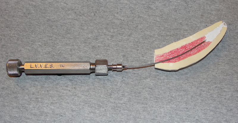
Dental Wax Technique to Pack Alveoli Following Extraction
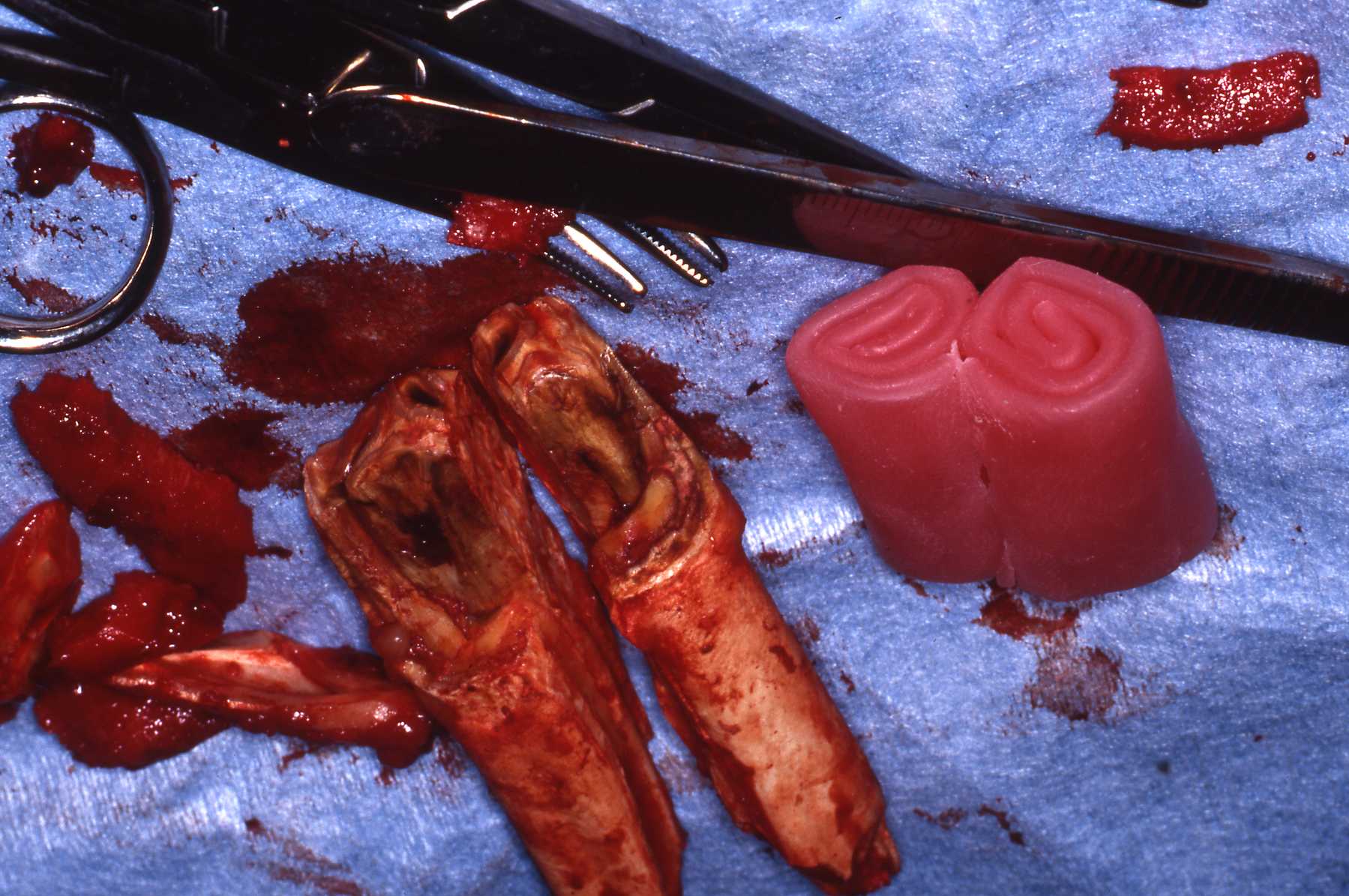
The author began to use pink dental sheet wax to pack dental alveoli following extractions in equine and ruminant cases in the 1990s. I have since learned that numerous other practitioners of veterinary dentistry have learned to use this technique. Alveoli that were surgically exposed must be surgically closed with tensionless flaps following appropriate alveoplasty. Those that were "simple" extractions can be packed with a wax plug that is molded to be approximately one half the length of the extracted tooth. The diameter is sized to fit tight in the alveolus. It is pushed into the alveolus to be flush with the gingival border.
The wax plug is comprised of natural substances that will gradually melt and be swallowed. It will keep food debris from entering the alveolus and permit granulation tissue to form in the base of the alveolus. It will stay in place for a week or two. I have use this technique in many species over the years with no negative consequences. For equine and some zoo cases I have done a follow up exam in 10 days and replaced the wax plug with a shorter one. One example that warranted this was a warthog mandibular tusk case.
Extraction Of Feline Teeth
The extraction of feline teeth follows the same principles described for the dog and other veterinary patients. However, a common indication for extraction of teeth in the cat is dental resorptive lesions. These lesions have been named Feline Odontoclastic Resorptive Lesions or FORL. See Reference section Topic: FORL. Teeth with dental resorptive lesions often are ankylosed, making extraction techniques very difficult. Affected teeth are easily fractured with even the most careful technique. The small size of the teeth and their close proximity present challenges for all techniques. When the teeth are fractured during the extraction procedure or when the crown has fractured spontaneously, the retained root can be retrieved by carefully reflecting a flap and removing the overlying bone to expose the root, or by pulverizing the root within the alveolus with a handpiece and dental bur. Although the risks associated with this latter technique have already been discussed, it is often the least traumatic and easiest method for root removal in the cat. Ankylosed roots that are not infected may be left in place. Simply smooth off the root to beneath the gingival level with the dental bur. The operator may consider reducing the root height and minor alveoplasty to permit closing the site with absorbable sutures.
When extracting teeth from small mammals such as the domestic cat size and smaller, the operator must be extremely cautious with forces applied to the mandible to avoid iatrogenic fractures. Again, the symphysis of carnivores is cartilaginous, and quite delicate in these small species.
Summary
Despite major advances in the practice of veterinary dentistry during the past decade. extractions are still the most commonly performed dental procedures. A sound understanding of tooth root anatomic features is important to proper extraction. Tooth extraction techniques using sound surgical principles will permit efficient procedures, minimize trauma and discomfort to the animal patient, and encourage rapid healing. The keys to success are controlled forces and patience during the extraction process.
This page:
Postoperative Care, Extraction of Feline Teeth, Summary
Page 1: Introduction, Indications, Morphology
Page 2: Techniques