Dr. John Scheels - Exotic Animal Dentistry Dr. John Scheels - Exotic Animal Dentistry
 
Dr. John Scheels - Exotic Animal Dentistry Dr. John Scheels - Exotic Animal Dentistry
Close ×
 

Banner Images and Descriptions

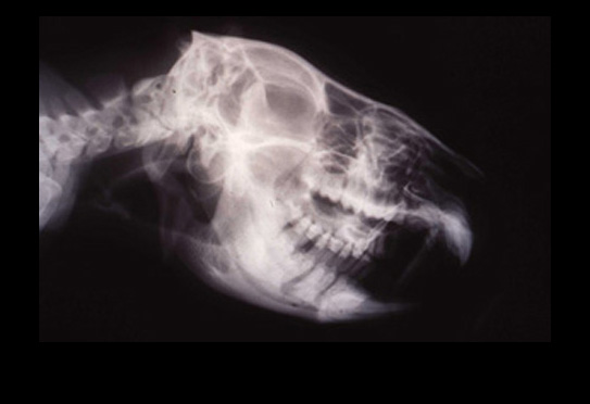
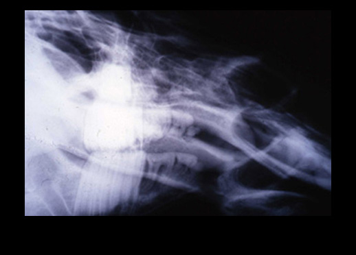
Koala Radiograph: Note interproximal bone loss due to periodontitis. Managed with periodic, every two months, “water pik” irrigation.
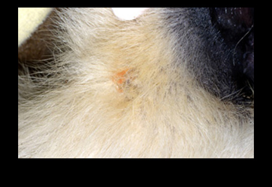
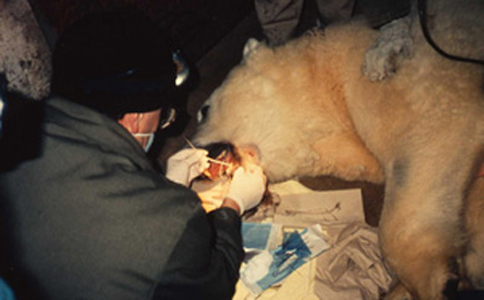
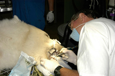
Polar Bear: Discolored hair due to chronic drainage tract from apex of abscessed mandibular canine. Extra oral apisectomy, retrograde endodontic treatment.
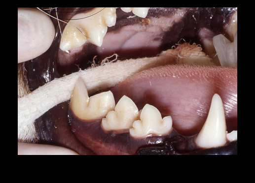
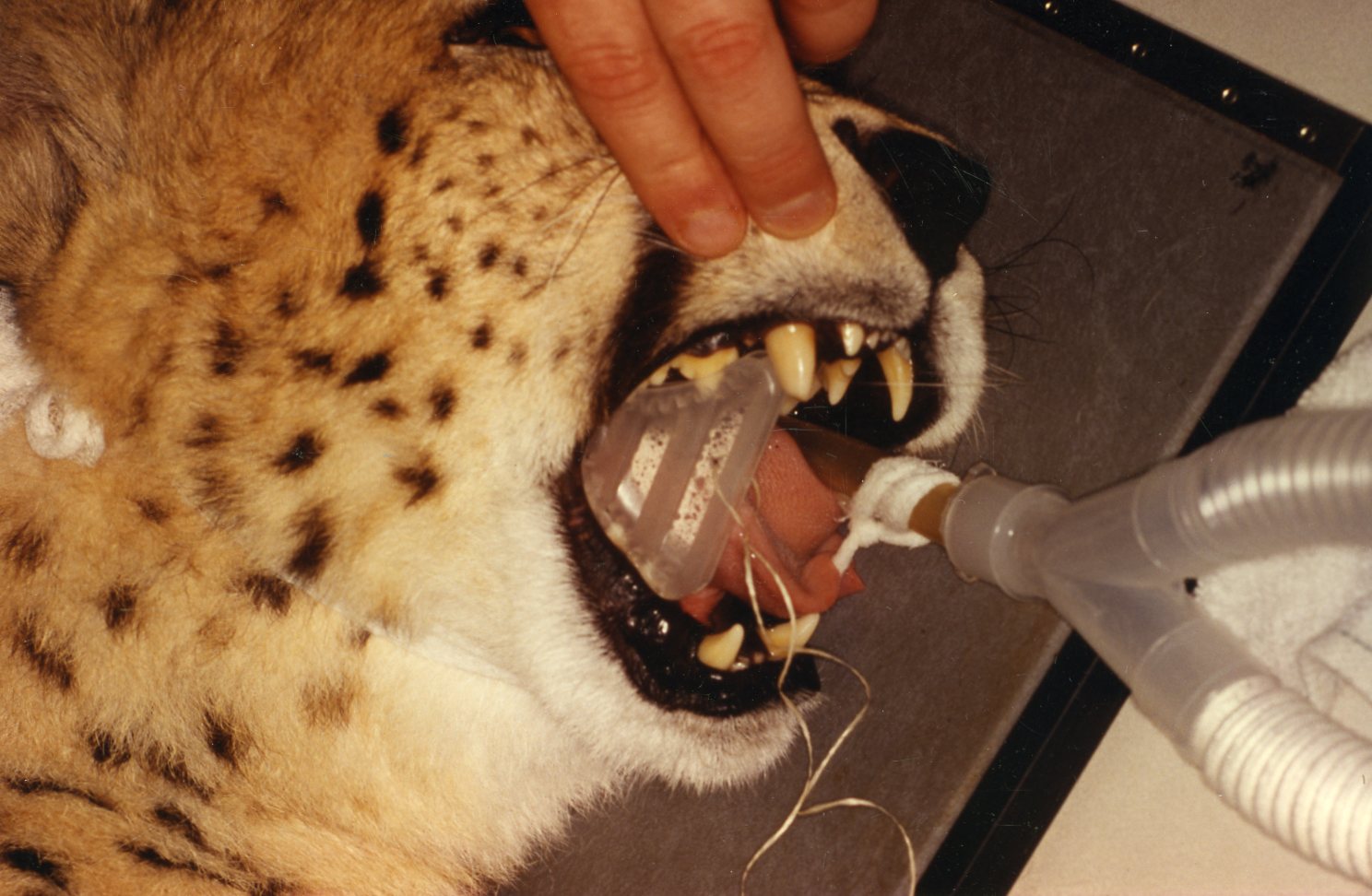
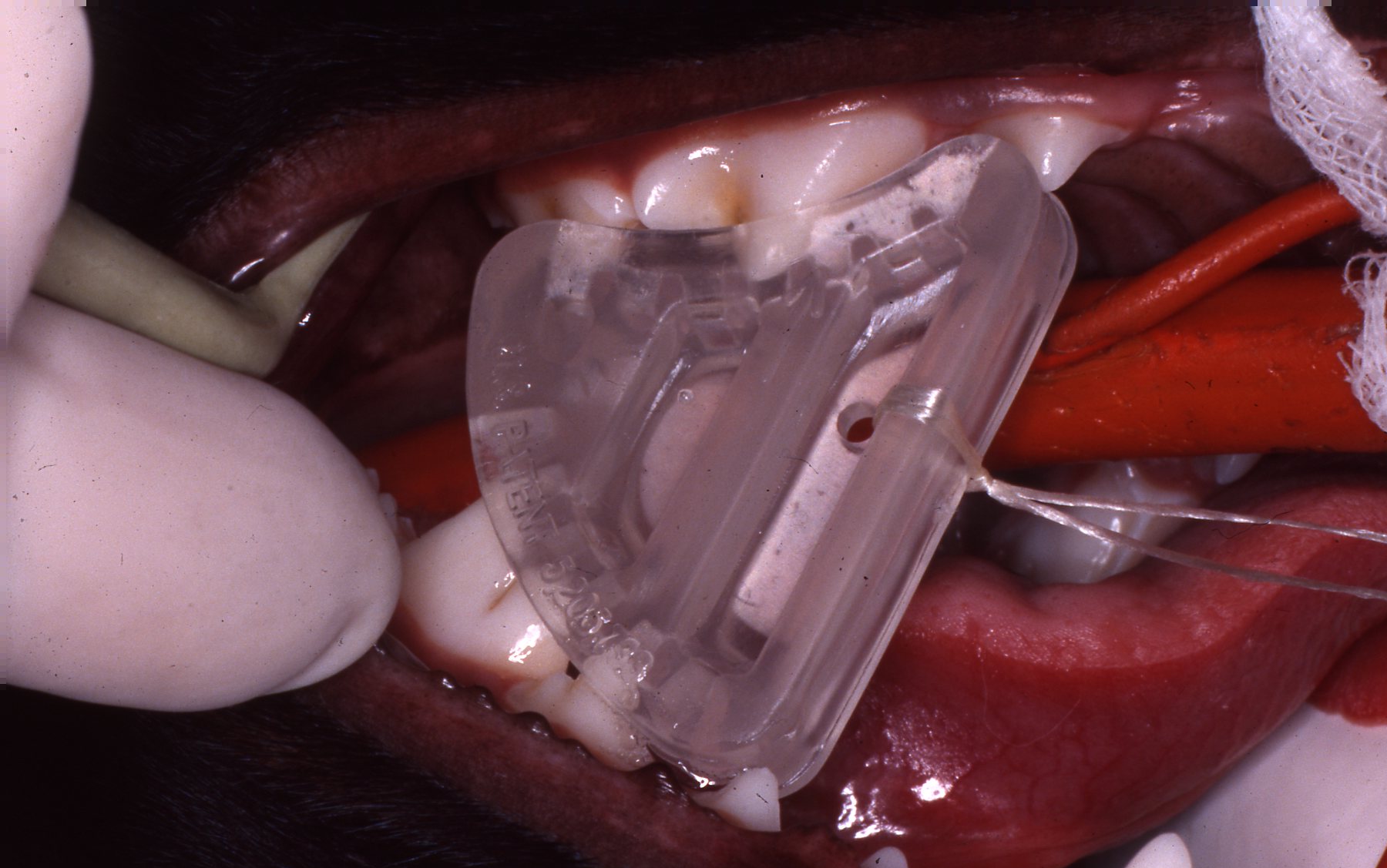
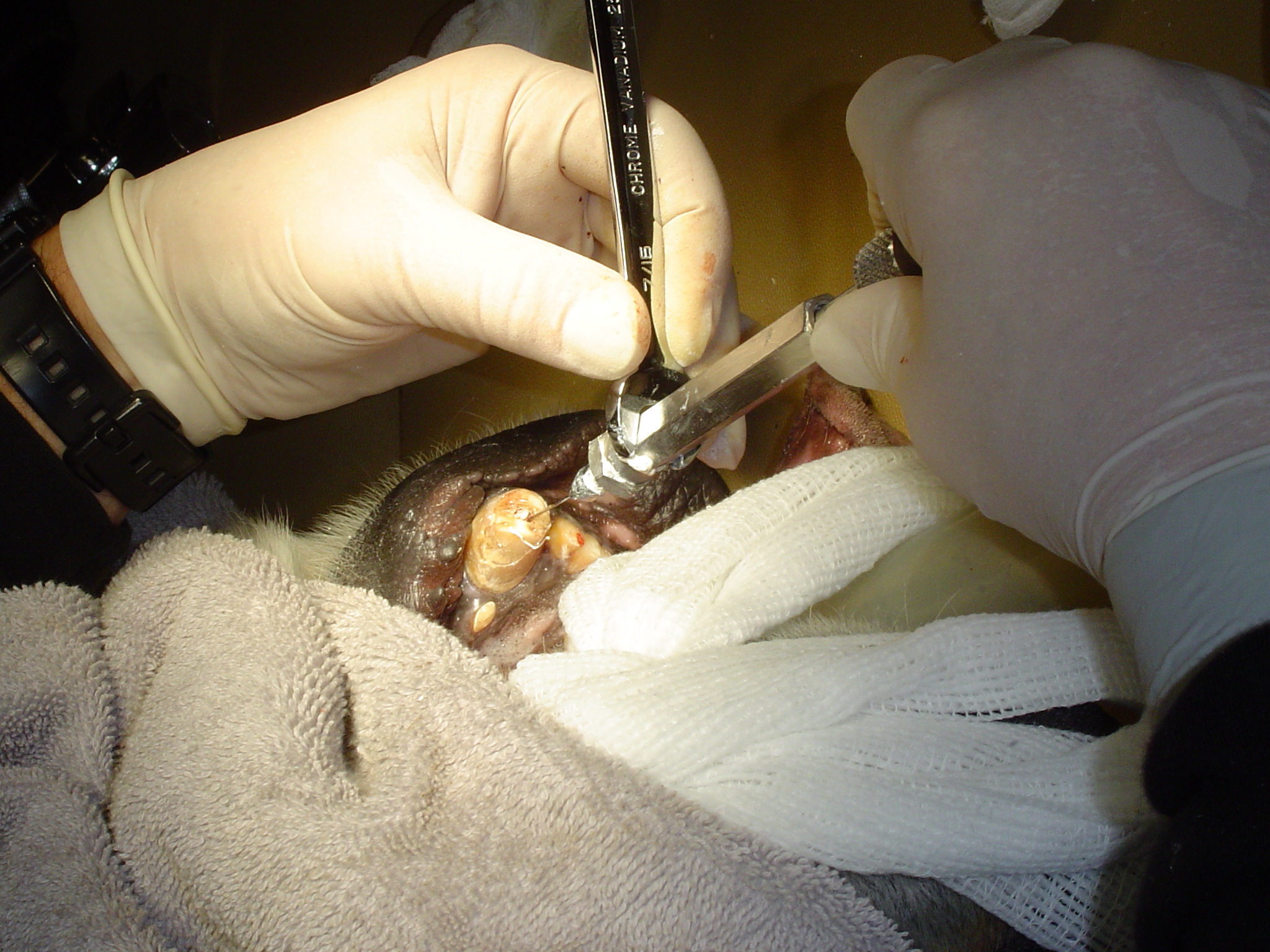
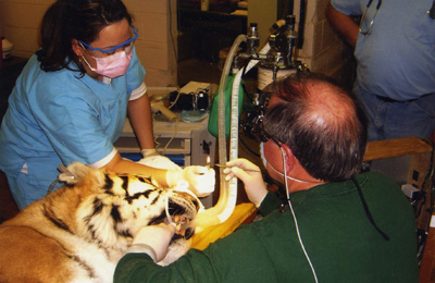
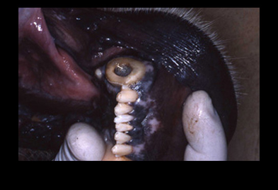
Cheetah: Overlapping mandibular teeth, malposed molar distal cusp protrudes into palatal fossa, causing “focal palatine erosion” lesion. Result of brachycephalic skull. Molar distal cusp was rounded off; radiographs necessary to determine proximity of pulp horn, to avoid pulp exposure. See: Focal Palatal Erosion in Captive and Free-Living Cheetahs (Acinonyx jubatus) and Other Felid Species, Zordan, Deem and Sanchez, Zoo Biology 30:1-8(2011)
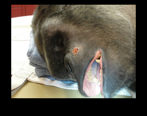
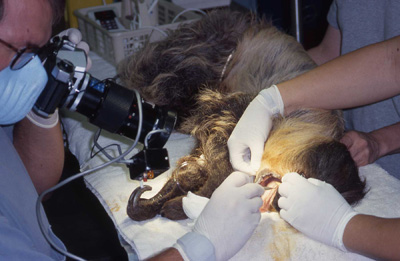
Gorilla: Extraoral chronic drainage tract from apex of abscessed maxillary canine. Endodontic treatment.
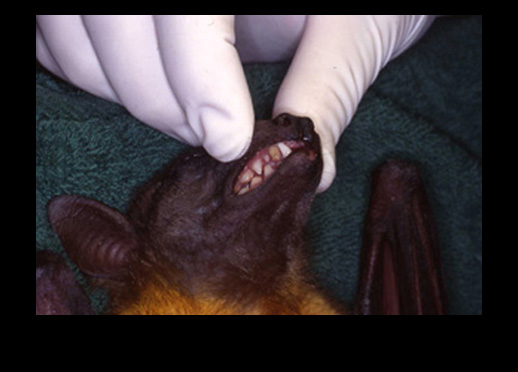
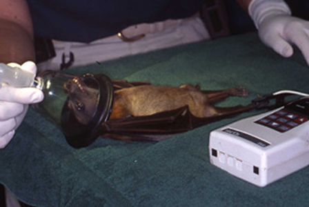
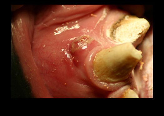
Fruit Bat: Darkened, abscessed maxillary canine. Extracted.
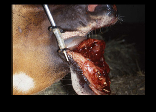
Impala: Bilaterally fractured mandible. Euthanasia because of management challenges if jaw was repaired, ruminant could not be tube fed during recovery.
Polar Bear: Fractured mandibular canine, pulp chamber filled with food debris. Extra oral apisectomy, retrograde endodontic treatment.
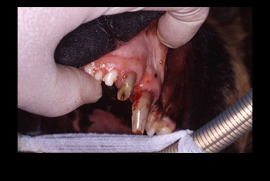
African Wild Dog: Fractured maxillary canine and incisor. Both extracted.
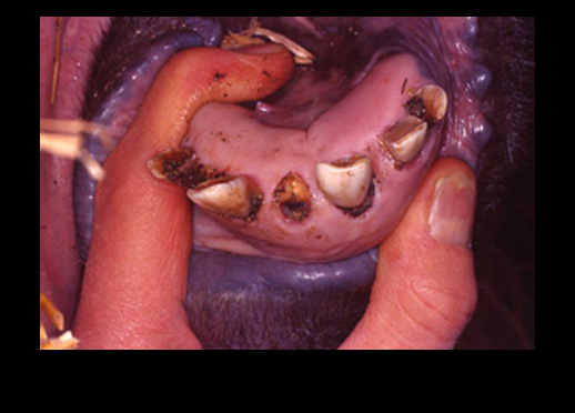
Warthog Radiograph: Avulsed mandibular “tusk”. Alveolus irrigated, filled two thirds length with wax plug to stop food impaction. Was replaced two weeks post op. Note very large maxillary and mandibular molars. Radiolucencies around first and second molars due to being pushed forward by “migrating” third molars.
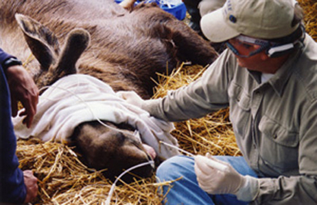
Moose: Fractured mandibular incisor. Extracted with luxators. No incisions necessary. Alveolus filled with two thirds length wax plug to avoid food impaction.
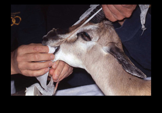
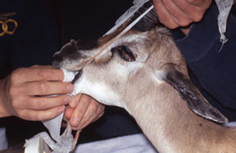
Spekes Gazelle: Swelling on mandible due to dental infection. Following tooth extraction, extra oral bone lesion debrided and packed with antibiotic impregnated beads. Drain placed when closed.
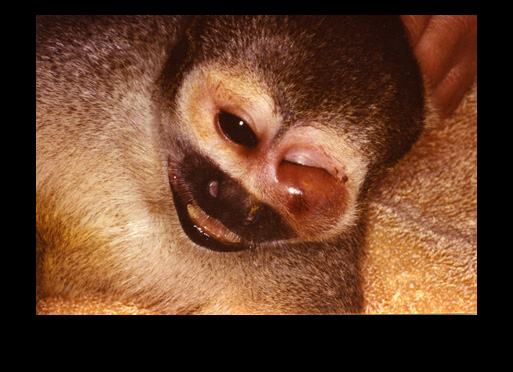
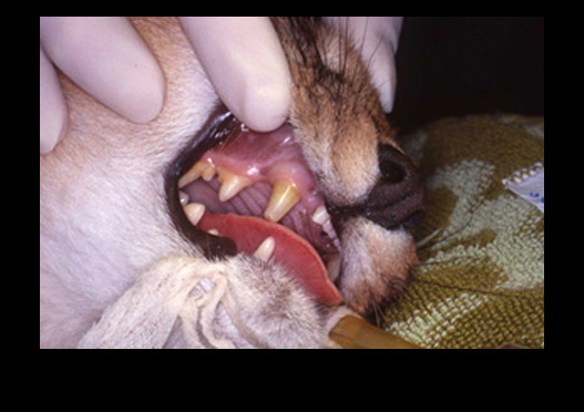
Squirrel monkey: Facial swelling due to abscessed maxillary canine. Extracted.
Caracal: Darkened maxillary canine. Oblique and extra oral apisectomy, retrograde endodontic treatment.
Bongo: Fractured mandible, within the symphysis. Part of the cartilaginous symphysis intact. Reduced with intermedullary pins, wired together.
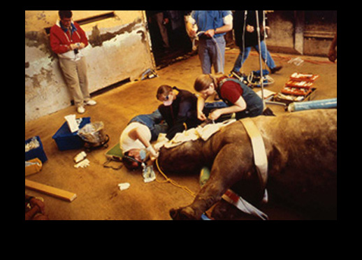
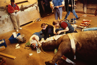
Black Rhino: Oral examination.
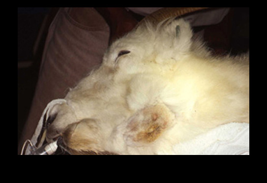
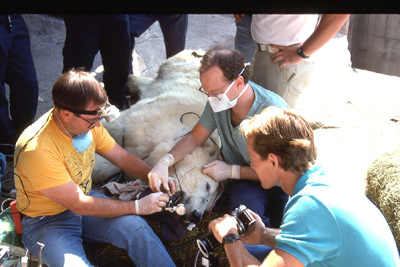
Dall sheep: Swollen mandible due to abscessed molar. Extra oral bone lesion debrided, and extra oral apisectomy, retrograde endodontic treatment.
Gorilla: Intra oral lesion, drainage tract, due to a pulpal abscessed canine. Endodontic treatment.

THE WEDGE®

Radiolucent Mouth Prop for Dogs and Cats

NOTICE: Veterinarians have used spring-loaded mouth "gags" in cats and dogs for many years. However, the spring-loaded devices are no longer recommended. A study published in THE VETERINARY JOURNAL (2014) showed that the spring-loaded "gags" generating constant force contributes to bulging of the soft tissues between the mandible and the tympanic bulla in cats. This force leads to the compression of the maxillary arteries as they course through the osseous structures. In cats the maxillary arteries are the main source of blood supply to the retinae and brain.

Reduction of the blood flow can result in temporary or permanent blindness and neurologic abnormalities. Spring-loaded "gags" constant force can also cause jaw muscle strain and injury to the temporomandibular joints.

It is recommended to use "static" mouth props such as the WEDGE. Be sure to not open the jaw to its maximum to avoid muscle strain and temporomandibular joint injury.

Dr. John Scheels - Exotic Animal Dentistry 
 

The WEDGE® is a one-piece, radiolucent mouth prop. The patented, anatomic design holds the carnivore mouth open during anesthesia by securely engaging the premolars and molars.

Dr. John Scheels - Exotic Animal Dentistry
PATENTED
 

The WEDGE®:

Dr. John Scheels - Exotic Animal Dentistry 
 

Developed and patented by Dr. John L. Scheels, dental consultant to the Milwaukee County Zoo, Adjunct Assistant Professor, University of Wisconsin-Madison School of Veterinary Medicine.

"The (Scheels) Veterinary mouth prop's biggest asset is its simplicity - open the mouth and stick it in!...it does not interfere with radiographic detail, can be ultrasounded, and autoclaved...can be used for dental and oral surgery procedures...is positioned within the mouth, unlike the spring loaded (extra-oral) devices which can be in the way of the operator and interfere with positioning the patient..."

~ Paul E. Howard, D.V.M.,

Vermont Veterinary Surgical Center, Burlington, Vermont.

Order The WEDGE® from any of these veterinary equipment suppliers:


L.V.V.E.S

Large Volume Veterinary Endodontic Syringe™

Click here for more details
Dr. John Scheels - Exotic Animal Dentistry 

An endodontic syringe developed by Dr. John Scheels specifically for veterinary use in all species for complete and consistent obturation of root canals over 30mm long or with large pulp chambers. It permits the positive deposition of endodontic sealer and filler pastes at the apex of these long teeth. NO SPECIAL NEEDLES are required as it may be used with any standard size hub. Plastic, metal, threaded or non-threaded needle hubs will seal well on the tapered syringe nipple.

Purchase from Shipp's

 
Dr. John Scheels - Exotic Animal Dentistry