


Small Patients
Zoo collections can include hundreds species from the entire animal kingdom, including reptiles, birds, fish and insects! The classic marquee zoo mammals include lions, tigers, gorillas, polar bears, elephants, and giraffes. However, there are mammals in zoos that are smaller and to most visitors may be considered less dramatic. They are certainly equally important species, serving as ambassadors for their wild counterparts, sustaining their genetics and helping to provide education about our wild world. The zoo staff and veterinarians provide the same optimum efforts to keep these smaller species happy and healthy. Often their smaller size alone presents medical management challenges.
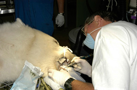
From drawing blood from tiny vessels, to intubating these diminuitive animals, establishing general anesthesia is often a significant challenge. For many cases that I’ve been involved with over the years, intubation was attempted by experienced veterinarians and zoo vet technicians, but after multiple tries it just couldn’t be accomplished. After attempts having taken too long, we continued using just injectable sedation/anesthesia and sometimes supplementing with anesthestic gas mask or cone held over the patients nose. This situation is of course not desirable but it is at least an option when dealing with a significant issue. It requires extremely close coordination by the veterinarians and veterinary technicians along with the dental operator.
Often the procedure is interrupted multiple times because the patient becomes too light to treat. When the anesthesia is resumed to a safe level we resume the procedure. We accomplished what was necessary in many such instances. In these situations prompt examination and evaluation of the animals dental condition is required. Decisions must be made what is most necessary to deal with and then the appropriate equipment put to use. Of course this once again emphasizes the need for thorough preparation for the particular patient, to anticipate what the patients situations may present and good communication between the veterinarian and dental operator.
Extractions are again the most common appropriate procedure. However, endodontics may be the best option for the patient to avoid a surgical extraction of a canine for example. Extracting a canine in a very small animal that requires a surgical approach is very challenging because of their small size. At the typical zoo hospital there simply isn’t the wherewithal to do micro surgery well.
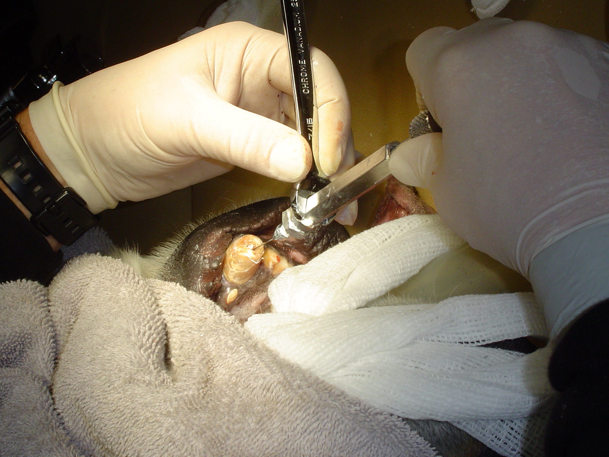
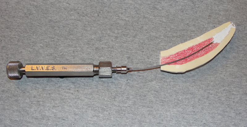
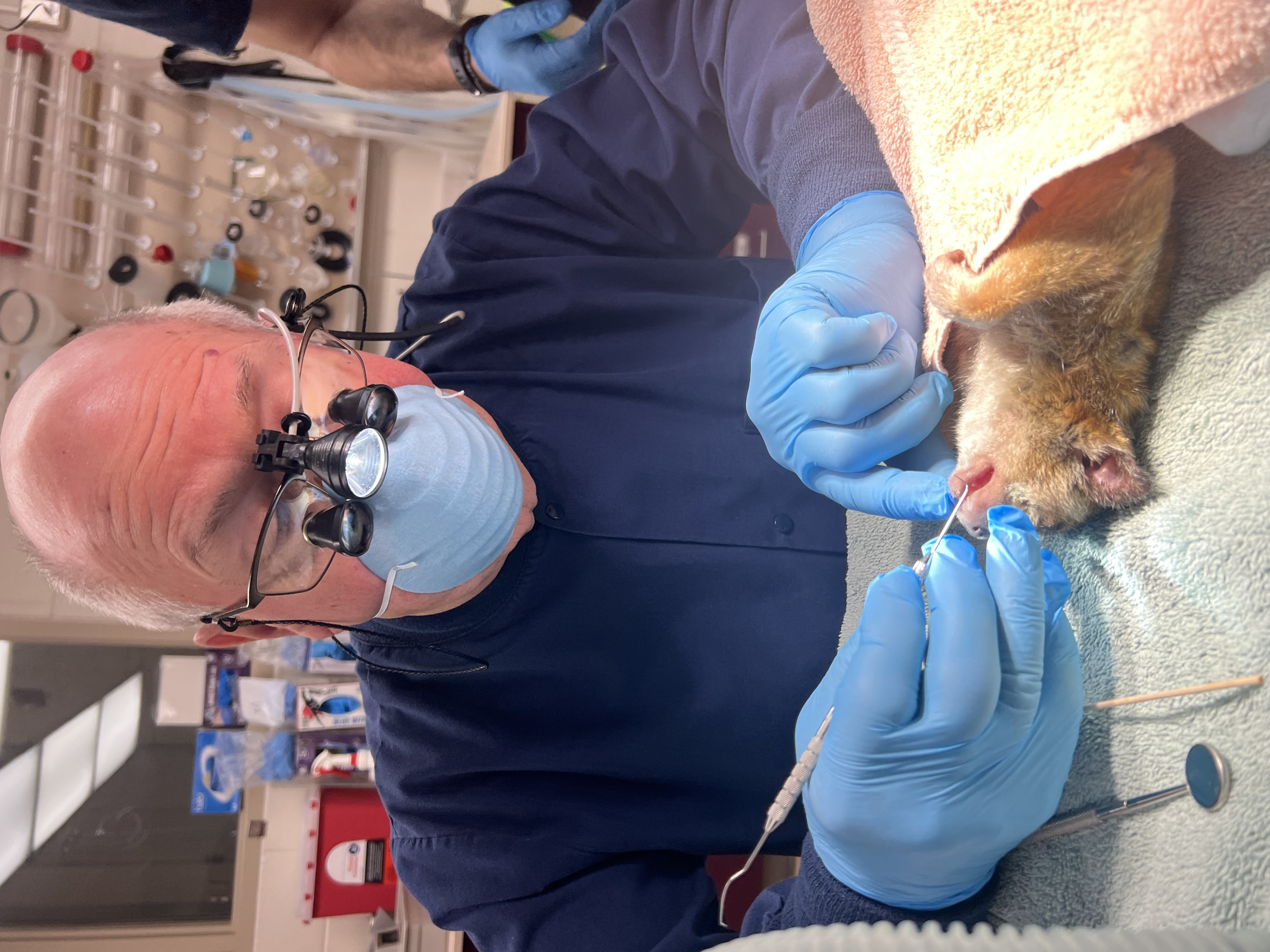
As pointed out in the Extraction Section, having appropriate sized instruments to deal with very small teeth is very important. Currently there are much smaller instruments available than when I first began treating animals in the 1980s. For example, tiny curved luxators are available from various instrument providers. See photo of the Cislak curved luxators that we use. One is concave on the outside of the curve, the other on the inside of the curve. These, among others are excellent, useful instruments. However, being so small, only 1.5 mm. wide, they are quite delicate and become dull quickly. Just as for the human sized dental luxators they must be maintained by resharpening. I do this by using appropriate sized dental lab carbide and stone burs. Modified hemostats in the appropriate size have been very useful. For example, I use a no. 8 carbide round dental bur to create a groove in the center of the beaks of the hemostat. This provides a space for the tooth to be grasped.
Often these small animals have heavy calculus deposits. The available dental scalers must be handled very carefully when removing these deposits. Pressure can be applied carefully by stabilizing the teeth from behind with a finger. For the tiny Galago with mandibular comb incisors the safest way to remove the heavy deposits is by gently pinching the incisors front and back to loosen the calculus. Note that many species have these seemingly mobile incisors that at first may appear to be mobile due to periodontal disease but actually are an adaptation for cleaning their hair coats by combing it. Mandibular comb incisors are found in numerous species, including various antelopes for example.
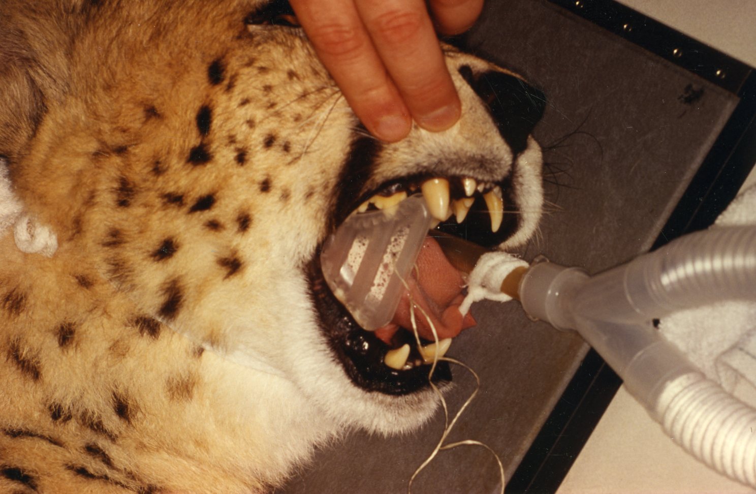
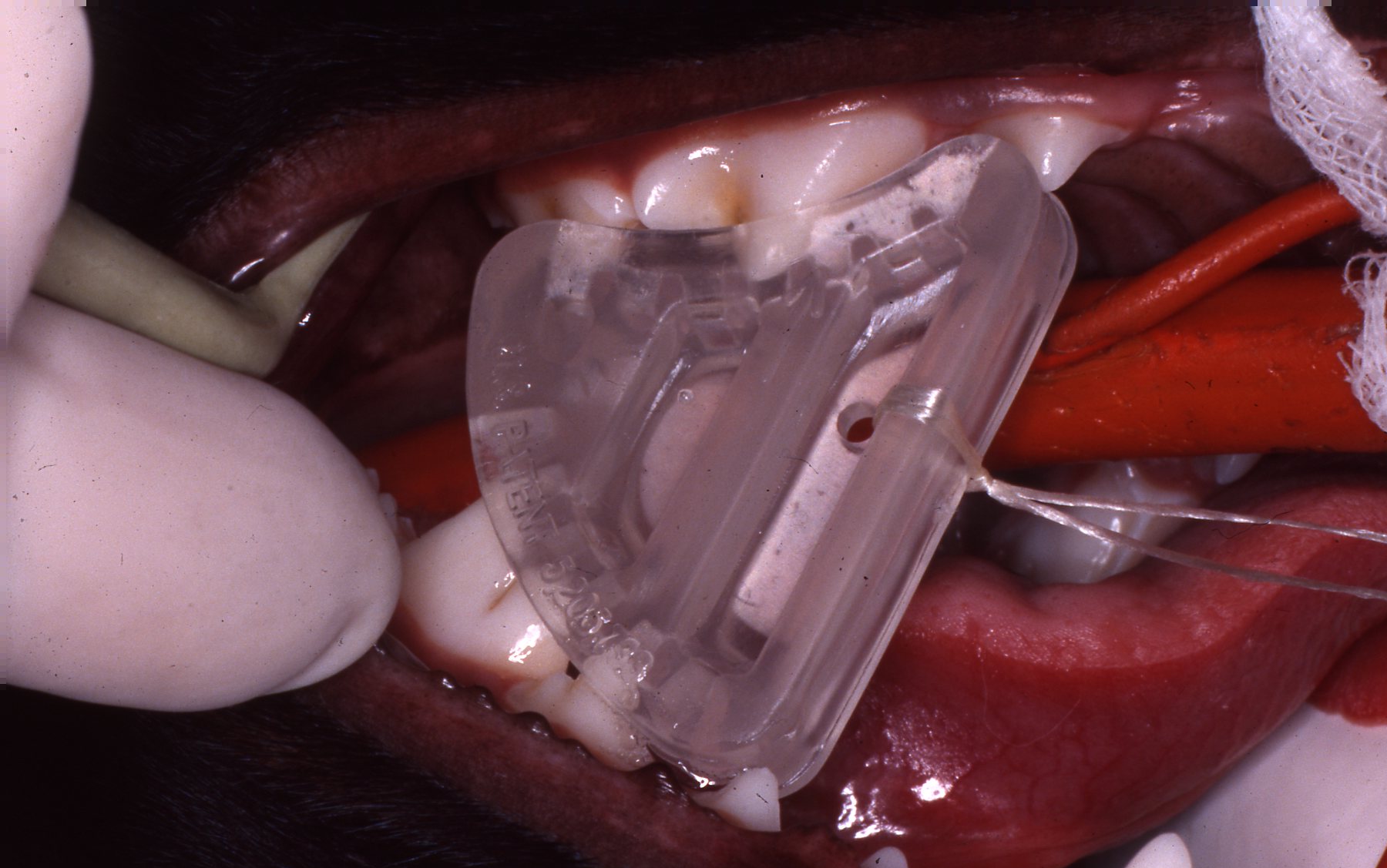
 Small animals present challenges due to small size
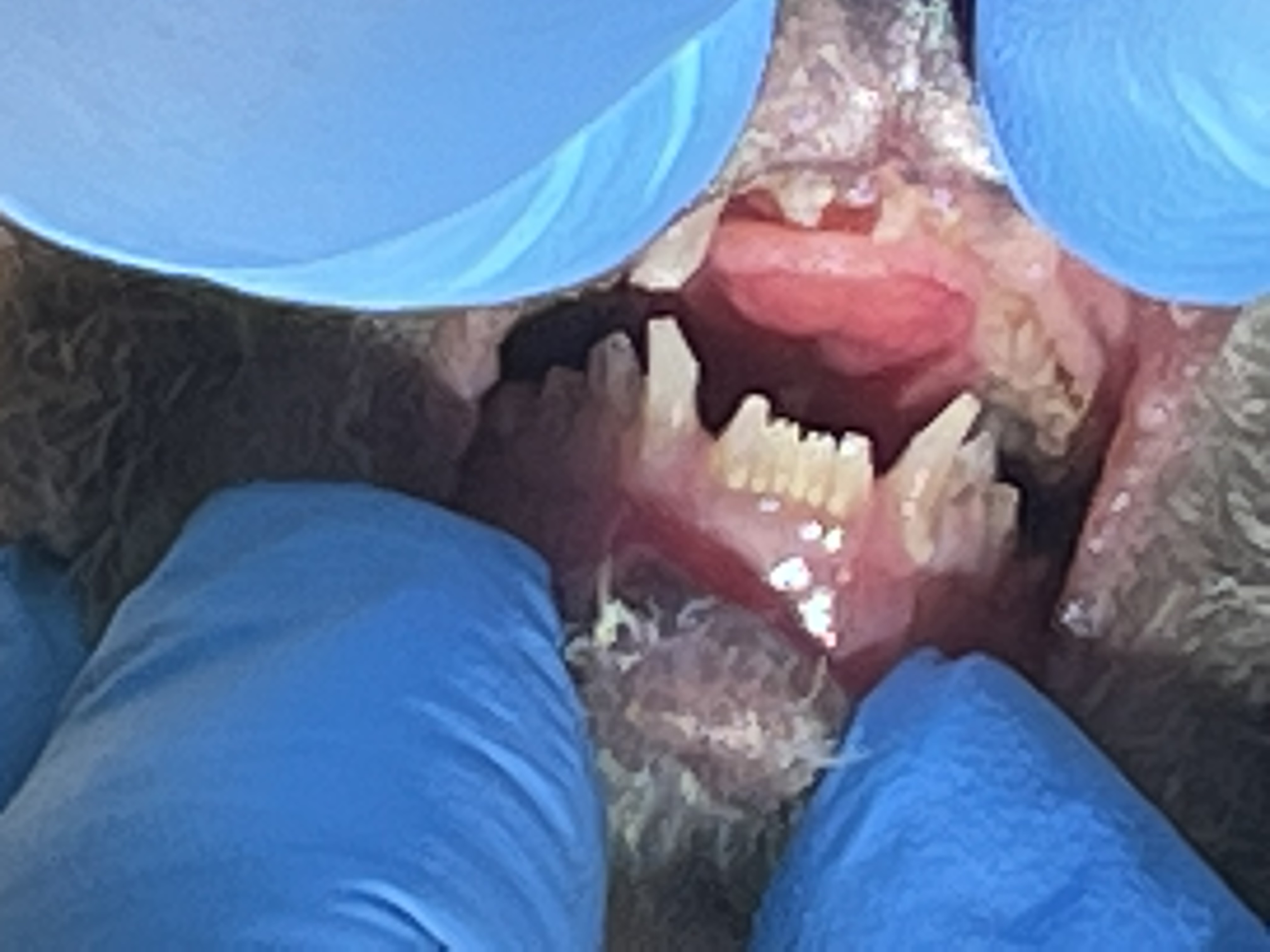 Calculus on small loris incisors
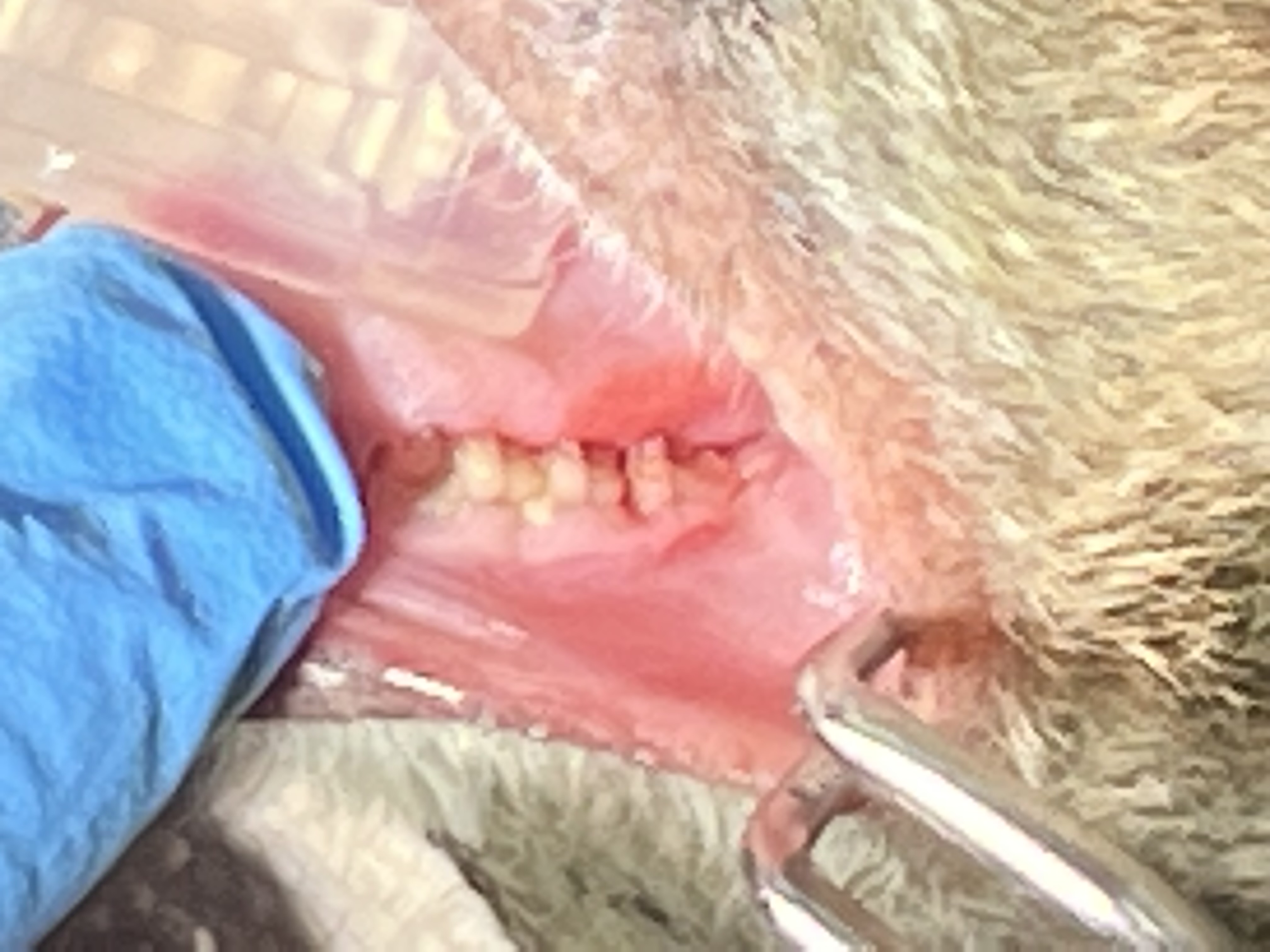 Deep distal periodontal pocket third molar
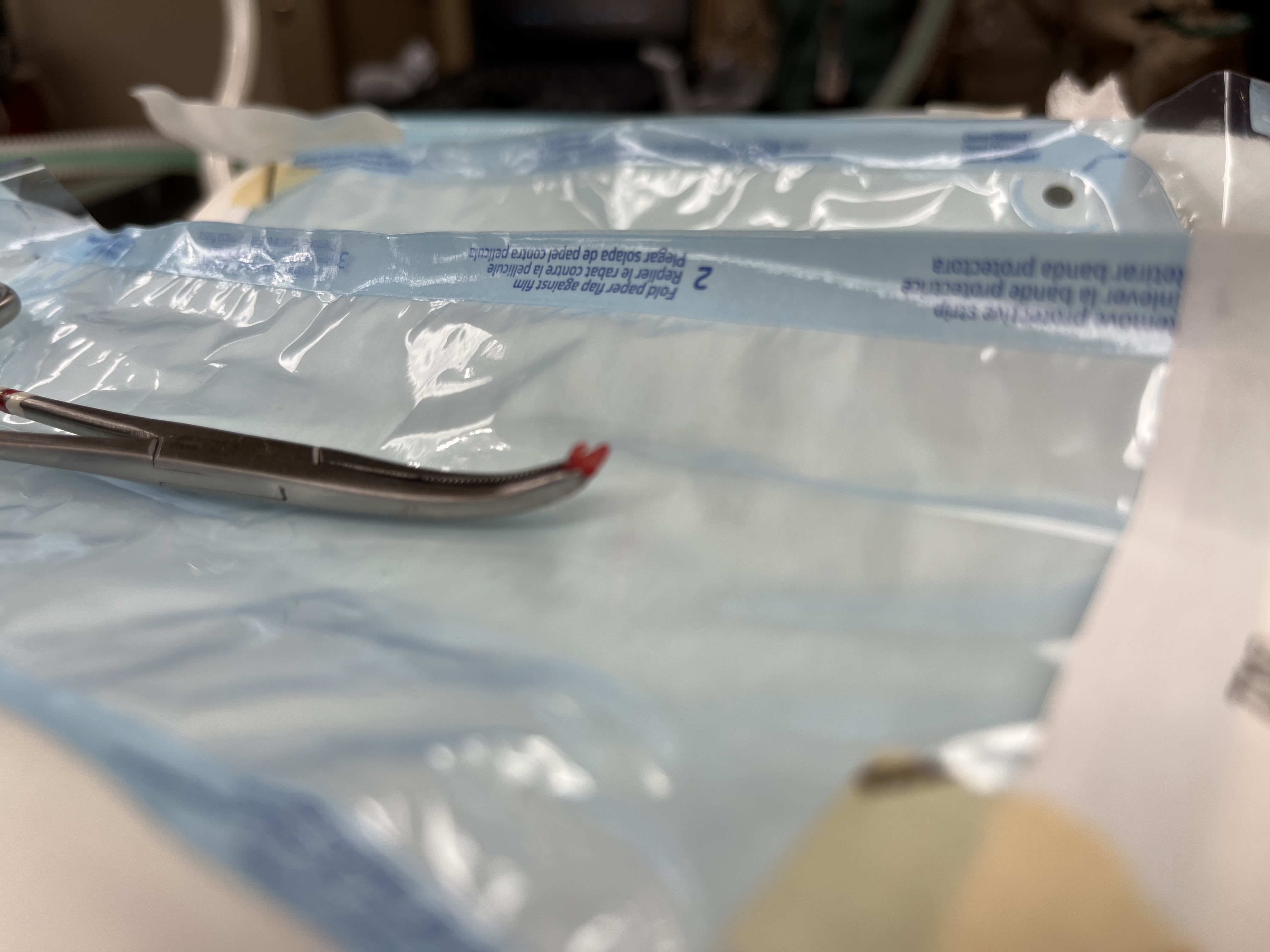 3mm long tooth
Galago, Kirby
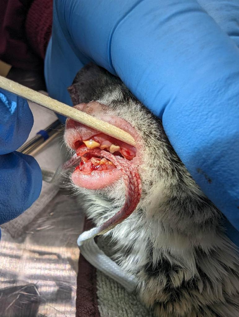 Galago heavy calculus
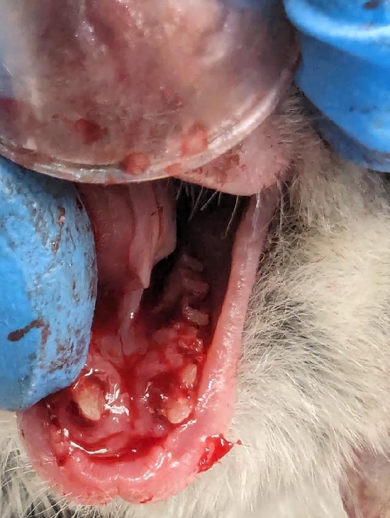 Deep lingual periodontal pocket first molar
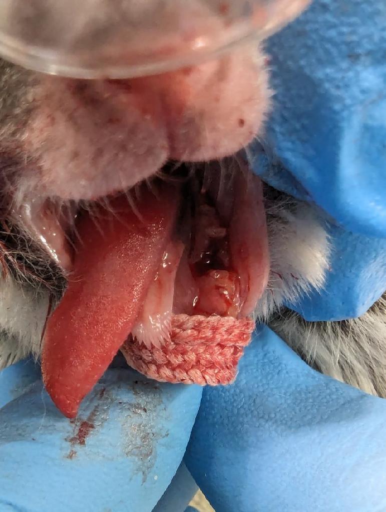 Molar extraction site
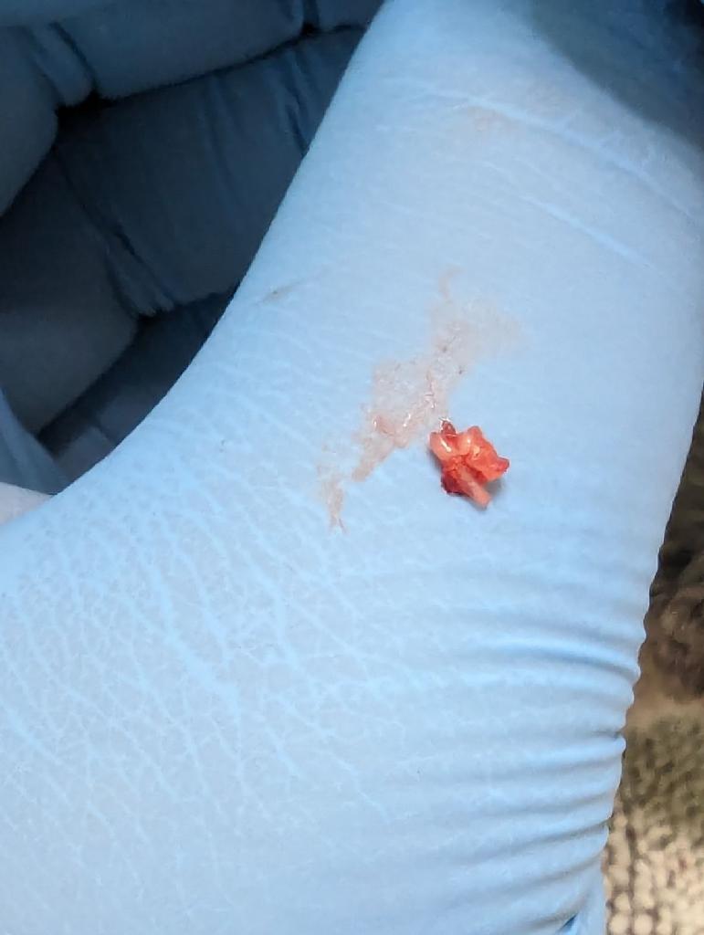 Extracted molar 3mm long and Cislak luxators
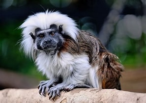
Cotton Top Tamarin
A 2022 Cotton Top Tamarin maxillary canine endodontic procedure is an example of successful endo case that avoided a surgical extraction for this very small patient. A working length radiograph revealed with file in place at 11 mm. showed that the file had gone beyond the apex. 8mm. was chosen as the proper working length and the endo procedure was completed. See fill radiograph.
 Cotton top tamarin
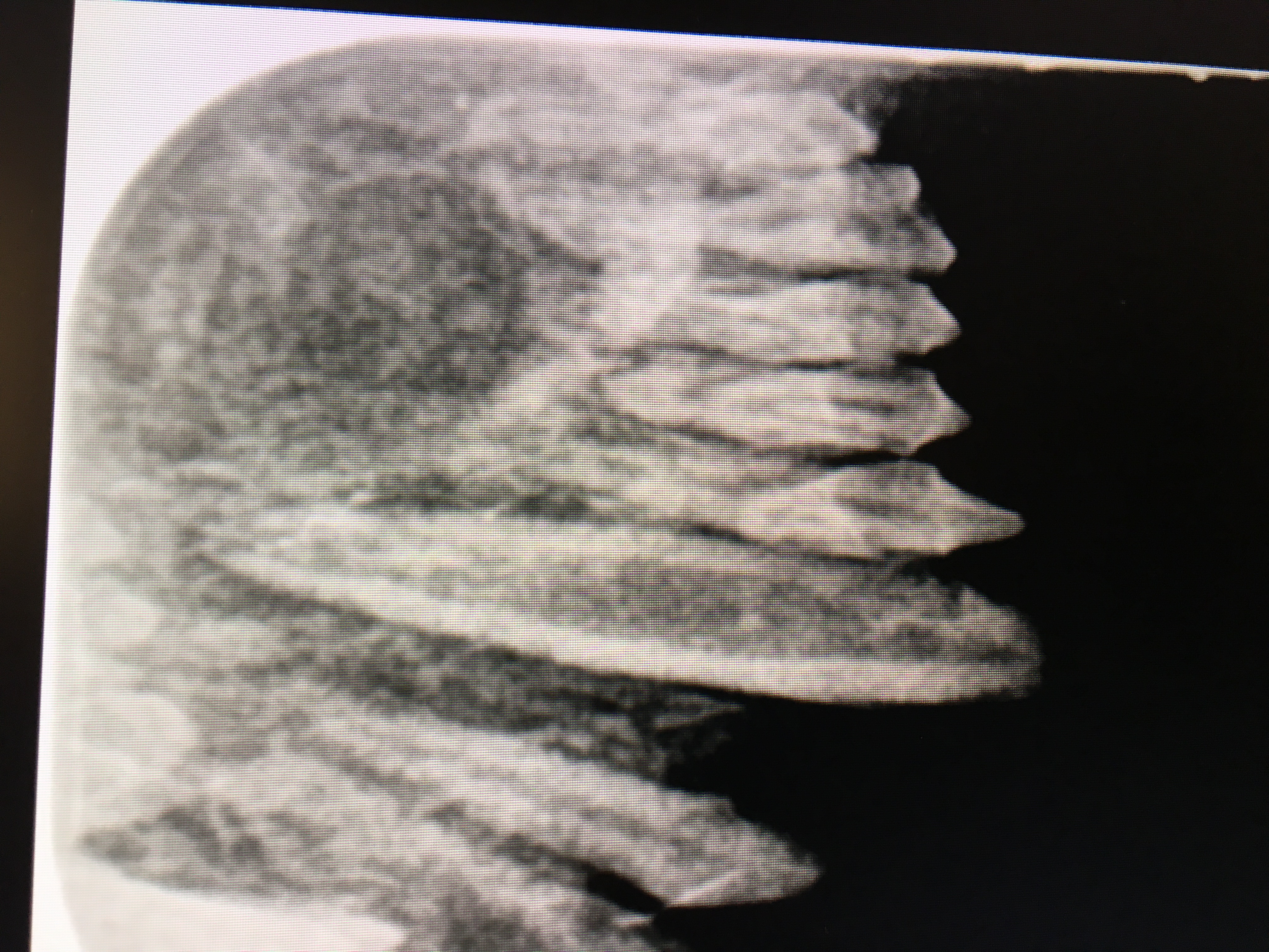 Preop radiograph of cotton top tamarin abscessed canine
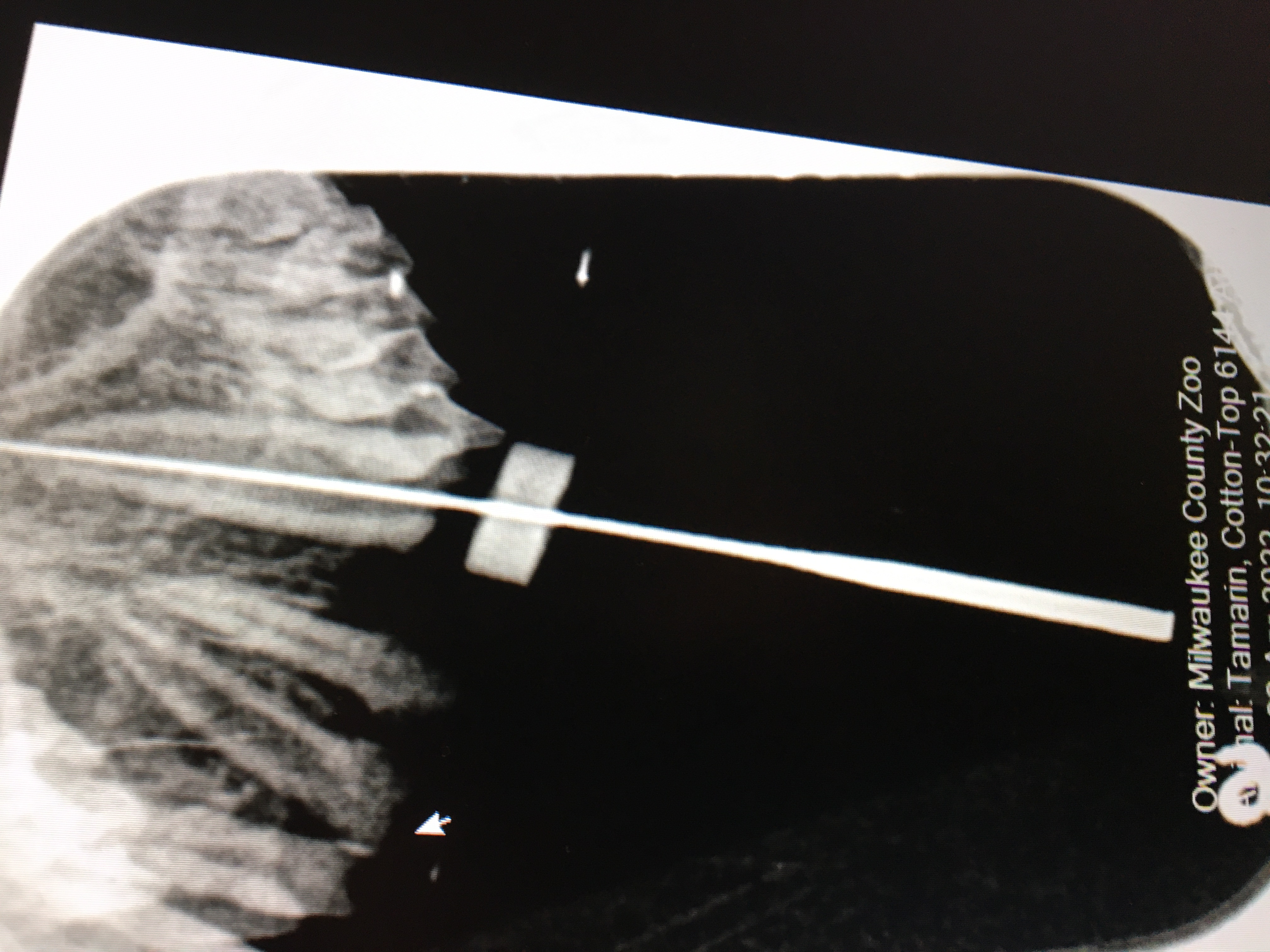 Working length radiograph 11mm
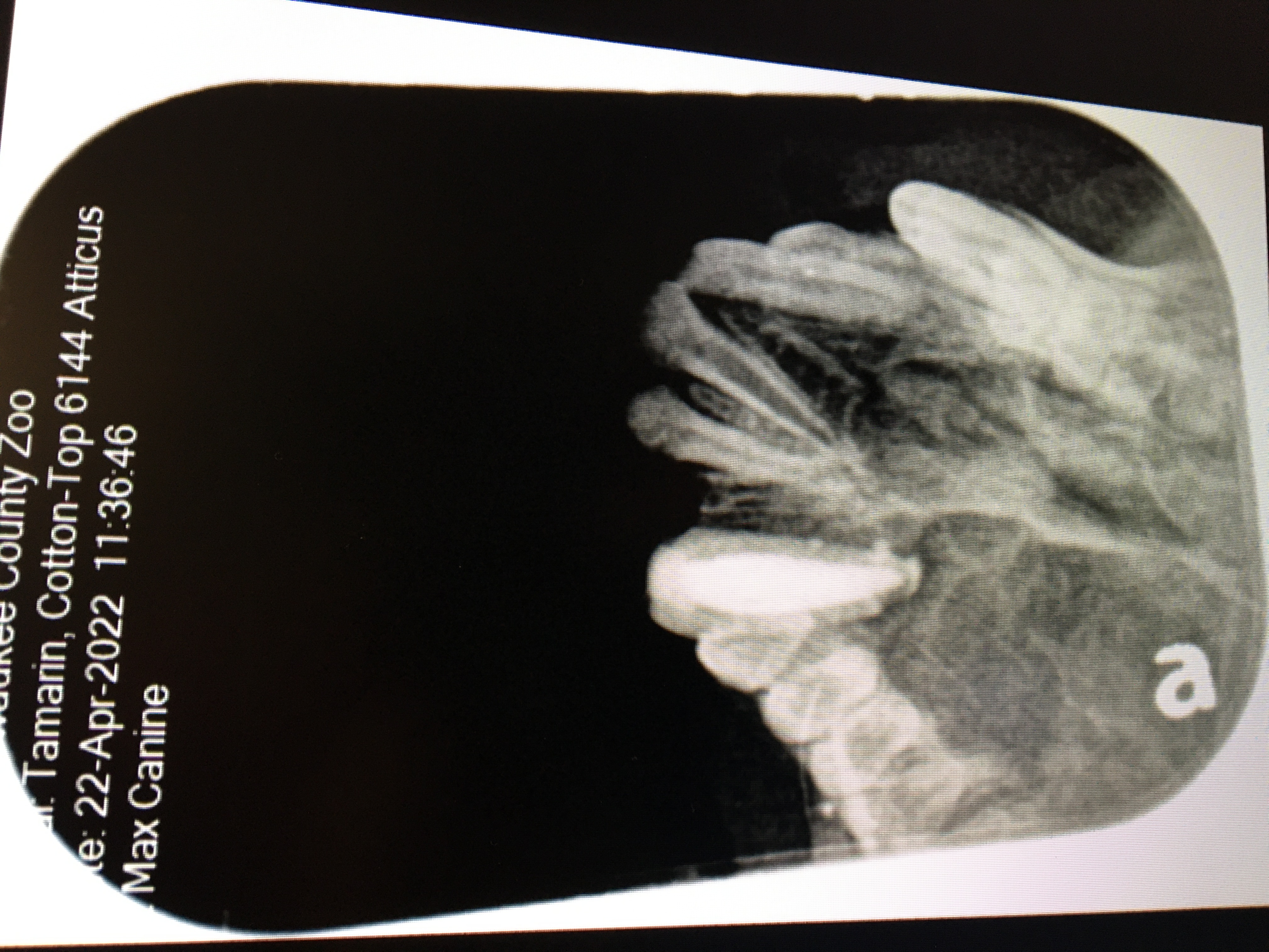 Endo fill at 8mm